

 Join
Join
Basal Cell vs. Squamous Cell Carcinoma: Differences and Pictures


Key Takeaways
- Basal cell carcinoma (BCC) and squamous cell carcinoma (SCC) are the two most common types of skin cancer, and while both develop on the outer layer of the skin, they grow and spread in different ways. View all takeaways
Sign up for free to view the full article summary.
Continue with EmailIf you’ve noticed a new sore or bump, you may be wondering if it could be skin cancer. The two most common types are basal cell carcinoma (BCC) and squamous cell carcinoma (SCC). While both types develop on the outer layer of skin, they have some key differences.
Recognizing the signs of BCC and SCC can help you get an early diagnosis and a better outlook. It can be challenging to tell them apart, so don’t hesitate to see your healthcare provider if you notice any skin changes. These skin cancers spread in different ways and require different types of treatment.
In this article, we’ll cover the differences between BCC and SCC, including how they form, what they look like, how they’re treated, and what to expect.
How BCC and SCC Develop and Spread
BCC and SCC are common types of skin cancer that form in the outer layer of the skin called the epidermis. Together, they make up 95 percent of all skin cancers.
BCC develops in the basal cells, which are located in the lowest layer of the epidermis (the outermost layer of the skin). Healthy basal cells produce new skin cells when old ones die and shed. BCC is the most common type of skin cancer and tends to grow slowly. It rarely spreads to distant areas of the body.
SCC develops in squamous cells, which are located closer to the skin’s surface and make up most of the epidermis. SCC is usually considered more aggressive than BCC. It tends to grow faster than BCC and is more likely to spread to nearby tissues, such as the lymph nodes. SCC may spread to distant areas of the body as well.
Both BCC and SCC are most likely to develop on areas of the skin that receive the most sunlight. That’s because the sun’s ultraviolet (UV) rays damage skin cells, and this damage can lead to cancer. Common areas for skin cancer to develop include the scalp, face, ears, and neck. SCC often develops on the lips, ears, and backs of hands.
Risk Factors for BCC and SCC
BCC and SCC share many of the same risk factors. The most significant risk factor for both types of skin cancer is exposure to UV light. This can come from the sun or a tanning bed. UV rays are the main cause of skin damage that can lead to cancer. They damage the DNA inside skin cells, changing the way the cells grow and behave.
According to the American Cancer Society, other risk factors for BCC and SCC include:
- Light-colored skin
- Older age
- Male sex
- Radiation exposure
- History of skin cancer
- Weakened immune system
Having a severe skin injury or infection can raise the risk of SCC. That’s because SCC can form in open sores or scars. Undergoing psoralens and ultraviolet light (PUVA) treatments for psoriasis can also raise the risk of SCC.
Other risk factors for SCC include smoking and a history of human papillomavirus (HPV) infection. People who smoke are at risk of developing SCC on their lips. Smoking doesn’t affect BCC risk. The sores caused by certain types of HPV may be related to SCC.
Symptoms of BCC vs. SCC
BCC and SCC usually develop on areas of the skin that receive the most sun exposure. BCC is often seen on the scalp, face, ears, neck, shoulders, and back. Because BCC grows very slowly, it can be challenging to recognize the signs.
Signs of BCC may include:
- A pearly bump — A BCC lesion may look shiny or waxy. It’s usually the same color as the skin and may have visible blood vessels.
- A sore that doesn’t heal — A common sign of BCC is an open sore that sometimes bleeds but never fully heals.
- A flat scar — In some cases, BCC looks like a scar that is brown or the same color as the skin. It may feel firm.
- A raised patch — BCC may cause a raised, crusted patch of skin that has a raised border.
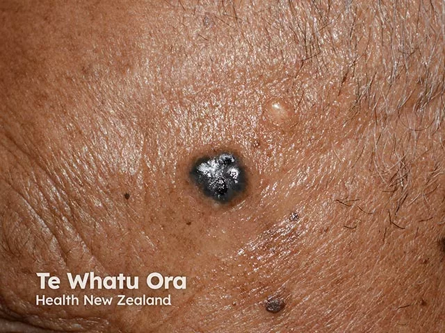
Pigmented basal cell carcinoma can look like a shiny, dark, raised bump on darker skin. (CC BY-NC-ND 4.0/DermNet)
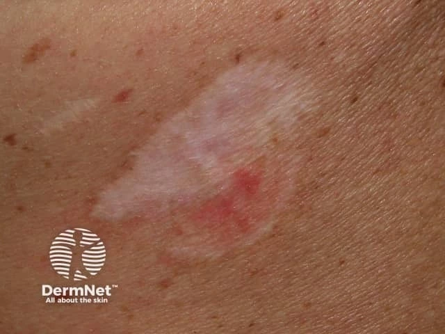
Morpheaform basal cell carcinoma can look like a pale, scarlike patch with subtle pink areas. (CC BY-NC-ND 4.0/DermNet)
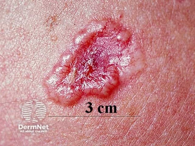
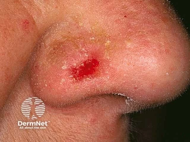
BCC appearance can vary by skin color. In people with lighter skin tones, the growth may appear red or pink. On darker skin tones, it may look blue, brown, black, or gray.
In most cases, BCC lesions don’t cause pain or itching. As they progress, the growths may start to break open, bleed, and crust over. Some people may develop pain, numbness, itching, sensitivity, or a pins-and-needles feeling as BCC grows.
Like BCC, SCC develops on areas of skin with the most sun exposure. It can also form on areas of skin irritation, such as a scar.
Signs of SCC may include:
- A scaly patch — A common sign of SCC is a crusted patch of skin that bleeds easily.
- A firm nodule — SCC may appear as a firm, red nodule that feels solid and rough.
- A sore that doesn’t heal — Like BCC, SCC can develop as a sore that never heals.
- A wartlike growth — SCC may look like a wart that is crusted and sometimes bleeds.
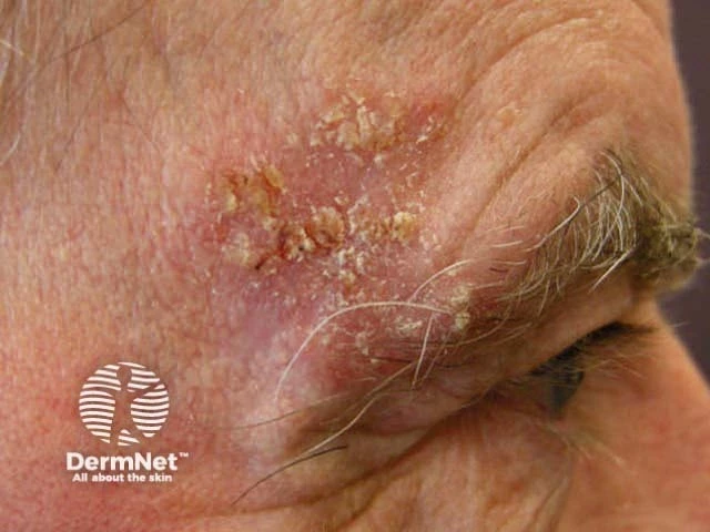
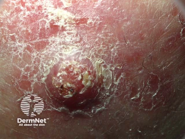
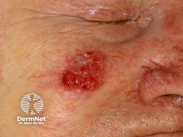
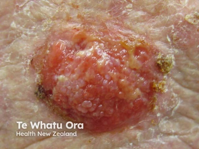
SCC may cause additional symptoms, including pain, itching, numbness, stinging, burning, or a pins-and-needles feeling. The pain usually gets worse as SCC progresses.
Treatment Options for BCC and SCC
Fortunately, both BCC and SCC are very treatable, especially when caught early. While BCC is usually less aggressive than SCC, both types of skin cancer can cause significant damage if left untreated. Your treatment plan will depend on several factors, including the type of skin cancer, where it’s located, how advanced it is, and your overall health.
The goal of BCC treatment is to remove the entire cancerous growth. Because BCC grows very slowly, it’s often possible to remove it completely. The goal of SCC treatment is usually to remove and destroy any cancer cells before they spread any further.
Surgery
When possible, your healthcare team will recommend surgery to remove skin cancer. When BCC and SCC haven’t spread to other tissues or areas of the body, surgery can be used to remove all cancer cells and cure the disease.
Surgical options for local BCC and SCC include:
- Excisional surgery — A surgeon removes the cancerous growth along with a margin of normal tissue.
- Mohs surgery — A specialized surgeon removes the cancerous growth layer by layer, examining the cells under a microscope to determine when all cancer cells have been cleared.
Excisional surgery is usually recommended when the entire tumor can be easily removed. Your healthcare team may recommend Mohs surgery if the tumor is large or in a sensitive area, such as close to the eye.
Topical Treatments
It’s rare for BCC to spread beyond the skin. That’s why it can often be treated with surgery or topical treatments applied to the skin. Your healthcare provider may recommend creams or gels that contain powerful medications to kill the cancer cells. These treatments are applied directly to the skin.
Another option to treat BCC is curettage and electrodesiccation. During this procedure, the dermatologist scrapes away cancer cells with a scooping tool called a curette. Then they destroy any remaining cancer cells with an electric needle.
Systemic Treatments
Some cases of SCC may require treatments that affect the entire body, known as systemic treatments. When SCC spreads beyond the skin, your healthcare team may recommend more aggressive treatments to destroy the cancer cells and prevent further spread.
Systemic treatments for SCC may include radiation therapy, chemotherapy, targeted therapy, and immunotherapy. These treatments are more common in SCC because it is more likely to spread.
Outlook for BCC vs. SCC
BCC and SCC are often curable when they’re found and treated early. BCC usually grows slowly and rarely spreads, but it can damage nearby skin and tissue if it isn’t treated.
SCC is also often curable when caught early, but it tends to grow faster than BCC and is more likely to spread to nearby lymph nodes or other parts of the body. Getting the right diagnosis helps your healthcare provider choose the best treatment and improve your outlook.
Join the Conversation
On MySkinCancerTeam, people share their experiences with nonmelanoma types of skin cancer, get advice, and find support from others who understand.
Have you been diagnosed with BCC or SCC? What were the first signs you noticed? Let others know in the comments below.
- Squamous Cell Carcinoma vs Basal Cell Carcinoma: Key Differences — American Oncology Institute
- Basal Cell Carcinoma vs Squamous Cell Carcinoma — Bare Dermatology
- Basal Cell Carcinoma and Squamous Cell Carcinoma: Comparison of High-Frequency Ultrasound and Pathology — Skin Research and Technology
- Basal and Squamous Cell Carcinoma Risk Factors — American Cancer Society
- Basal Cell Carcinoma: From Symptoms to Treatments — American Academy of Dermatology
- Basal Cell Carcinoma — DermNet
- Squamous Cell Carcinoma: From Symptoms to Treatments — American Academy of Dermatology
- Cutaneous Squamous Cell Carcinoma — DermNet
- Treating Basal and Squamous Cell Skin Cancer — American Cancer Society
Related
- How does skin cancer progress over time? See answer
- Who needs skin cancer screening? See answer
- Can skin cancer come back after treatment? See answer
- What does squamous cell carcinoma look like? See answer






 Your Privacy Choices

I just had a SCC taken off my left arm That was bigger than a quarter coin. I did it by the MOHS method. The surgeon said that she got it all. this is the 5th skin cancer I have had removed. I also lost my left kidney to cancer and have had two bladder tumors removed in the last 7 yrs.